

Lasers, Magnetic Stimulation and a Robotic Arm: How Researchers at HSE University Study the Brain
The Institute for Cognitive Neuroscience (ICN) at HSE University has recently added state-of-the-art laboratory equipment to its range of tools for studying brain function. The News Service visited the Institute to learn more about the uses of infrared lasers, optical tomography and a unique robotic arm, as well as why research into vascular tone is important, which parts of the brain can be stimulated to make people more generous, and how the Institute’s research can help treat diseases.
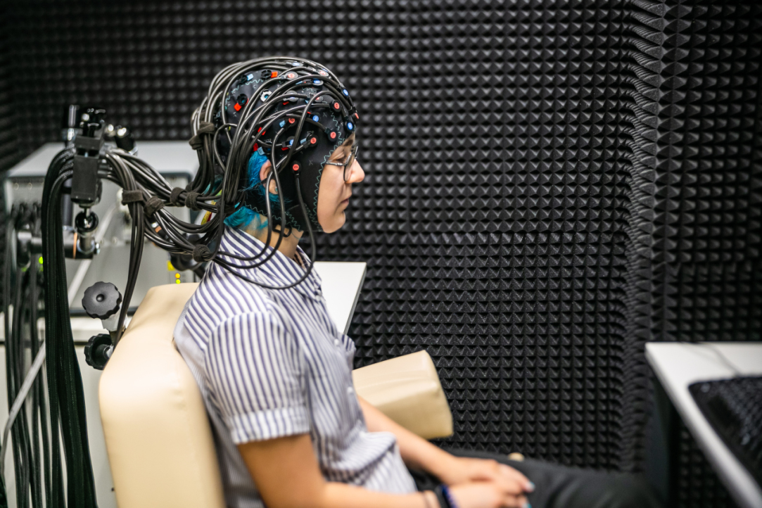
The Functional Optical Tomography laboratory looks like a medical room for EEG examinations. A comfortable chair stands next to a computer, which is connected to a ‘helmet’ made up of wires and dozens of sensors that envelop the test subject’s head. The room containing the optical tomograph itself is shielded by a ‘shell’ of special material that protects the device from external interference.
Aleksei Gorin, Junior Research Fellow at the Centre for Cognition and Decision Making at the ICN, told the HSE News Service how the process works. Special sensors conduct optical pulses from infrared lasers to detect differences in vascular tone in the veins and arteries. This in turn is used to measure oxygen absorption levels in various parts of the brain associated with logical problem solving and mental, sensory and motor activity. This allows researchers to study the brain's capabilities and its ability to respond to different tasks with more precision.
When our News Service reporter tested the equipment by reading a poem to himself, there was a drop in signal strength from the sensors monitoring the concentration of oxygenated blood. This, explained laboratory staff, reflects an increase in oxygen consumption in the brain.
The new equipment can also be used to study how the brain works when making certain decisions
The technology makes it possible to investigate certain kinds of brain activity and their impact on the brain's computational (and other) abilities. Optical tomography research also has medical applications, namely for the early diagnosis of heart and brain diseases.
In the absence of a perfect tool for studying the brain, a variety of experimental methods are required to get a clearer picture, Mr. Gorin explained. Researchers can conduct detailed behavioural research on the influence of various brain processes on human behaviour, and predict with a reasonable degree of accuracy which decisions an individual might make while affected by one process or another. ‘You can also give the brain a task and see how external factors affect how the brain works when performing decision-making tasks, sensory tasks or attention tasks. You can also learn more about how the brain works when someone is resting and relaxing’, he said.


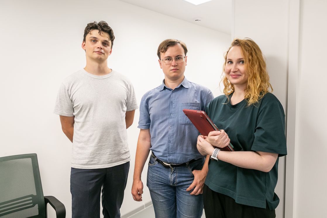
Mr. Gorin also said that the Laboratory is interested in conducting interdisciplinary research with economists and medical professionals. Over the last two years, the Laboratory’s staff has expanded to include other specialists, including neurologists and psychiatrists. This has helped broaden the scope of its brain research.
Tatiana Chernyakova, Research Assistant at the International Laboratory of Social Neurobiology, said that the Laboratory’s plans for the near future include neuromarketing research and two cognitive experiments with the new equipment, using a combination of MRI and optical tomography to obtain new data.
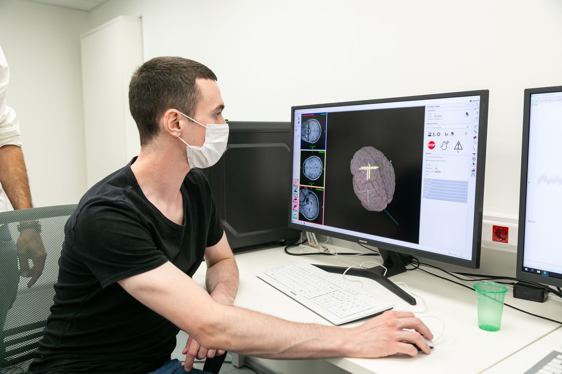
The Transcranial Magnetic Stimulation (TMS) laboratory has been overseeing the operation of a state-of-the-art stimulation device capable of activating parts of the brain. The device is installed next to a chair similar to one found in a dentist's office. A unique robotic arm (manufactured in France as there is currently no Russian equivalent) is placed above the head of a test subject and adapts to their movements. The accuracy of the robotic arm can be checked using a computer containing information about the area to be stimulated.
Matteo Feurra, Leading Research Fellow at the ICN Centre for Cognition and Decision Making, explains that if the subject makes any sudden movements for any reason (such as feeling unwell), the robotic arm deactivates the pulses and withdraws to a safe distance.


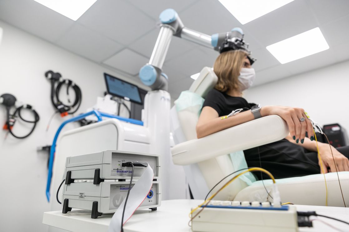

During the stimulation process, the robotic arm moves closer to the subject’s head, on which a reference bar has been placed to keep track of its position. The robot arm moves to match even the slightest movements of the head and sends an impulse to the target area of the brain. This induces the desired reaction, such as the contraction of muscles in the arm or fingers. While this result is visible in the test subject, the induced potential is also recorded and displayed by the computer.
Oksana Zinchenko, Research Fellow at the International Laboratory for Social Neurobiology, noted that stimulation has both research and medical applications. Once such use is the restoration of cells located around lesions or in the opposite hemisphere to an area affected by a stroke. Research Fellow Ainur Ragimova explained that transcranial magnetic stimulation can be used to treat depression and cervical dystonia, a disease that causes neck spasms, difficulty turning the neck and pain.
The TMS laboratory has a dedicated room for conducting complex research into brain stimulation and recording its effects. It is also shielded by a 'shell' for maximum signal clarity and isolation from external influences.
Areas of interest for this research include decision-making, social decisions, the speech areas of the brain, and memory. Ms. Zinchenko also explained that TMS is used to study the prefrontal cortex of the frontal lobe, which is responsible for selfish or prosocial motivations when deciding how to allocate resources—such as choosing whether or not to share points in a social game.
Using TMS to block the prefrontal cortex makes a person's behaviour more prosocial—they become more generous
Shutting down a group of neurons makes it possible to identify certain causal relationships. Researchers can see whether shutting down or stimulating individual areas disrupts or improves performance at a given task, and thus determine which areas of the brain are responsible for said tasks. These findings can then be used to build cognitive maps.
The laboratories and various research methods are available to specialists from different departments of the Institute for Cognitive Neurosciences, including foreign scientists such as Iiro Jääskeläinen, Academic Advisor of the International Laboratory of Social Neurobiology, Maria Del Carmen Herrojo-Ruiz, Leading Research Fellow at the ICN, Vladimir Djurdjevic, Research Fellow of the Centre for Cognition and Decision Making, Matteo Feurra and others.

Aleksei Gorin
Vladimir Djurdjevic
Tatiana Chernyakova
Maria Del Carmen Herrojo-Ruiz
See also:

Scientists Develop Bacterium-Sized Microlaser
An international team of researchers, including scientists from HSE University–St Petersburg, has developed microlasers that emit deep-ultraviolet light at a wavelength of 255 nanometres. The devices operate at room temperature, and the smallest of them measures just two micrometres in diameter—roughly the size of a bacterium. These microlasers could be used in sensors, spectroscopic systems, photonic chips, and communication devices. The paper has been published in Optics & Laser Technology.
Why Ice Cream Tastes Better in Hot Weather: HSE's Science Republic Takes Part in Popular Science Festival at VDNKh
In early June, the Science Republic initiative of the Centre for Student Academic Development presented popular science projects at the Science and Ice Cream Festival at VDNKh. On June 5, 13, and 14, HSE University ran an extensive programme of events, from lectures to intellectual games. The events took place at the BIOTECH Museum, Museum of Urban Economy of Moscow, and Smart City pavilions.
Wearable Device Data and Saliva Biomarkers Help Assess Stress Resilience
A team of scientists, including researchers from HSE University, has proposed a method for assessing stress resilience using physiological markers derived from wearable devices and saliva samples. The participants who adapted better to stress showed higher heart rate variability, higher zinc concentrations in saliva, and lower potassium levels. The findings were published in the Journal of Molecular Neuroscience.
HSE Researchers Propose New Method of Verbal Fluency Analysis for Early Detection of Cognitive Impairment
Researchers from the HSE Center for Language and Brain and the Mental Health Research Centre have proposed a new method of linguistic analysis that enables the distinction between normal and pathological ageing. Using this approach, they showed that patterns in patients’ word choices during verbal fluency tests allow clinicians to more accurately differentiate clinically significant impairments from subjective memory complaints. Incorporating this type of analysis into clinical practice could improve the accuracy of early dementia diagnosis. The results have been published in Applied Neuropsychology: Adult.
Scientists Reveal How Language Supports Complex Cognitive Processing in the Brain
Valeria Vinogradova, a researcher at HSE University, together with British colleagues, studied how language proficiency affects cognitive processing in deaf adults. The study showed that higher language proficiency—regardless of whether the language is signed or spoken—is associated with higher activity and stronger functional connectivity within the brain network responsible for cognitive task performance. The findings have been published in Cerebral Cortex.
Scientists Show That Peer Influence Can Be as Effective as Expert Advice
Eating habits can be shaped not only by the authority of medical experts but also through ordinary conversations among friends. Researchers at HSE University have shown that advice from peers to reduce sugar consumption is just as effective as advice from experts. The study's findings have been published in Frontiers in Nutrition.
HSE Scientists Uncover How Authoritativeness Shapes Trust
Researchers at the HSE Institute for Cognitive Neuroscience have studied how the brain responds to audio deepfakes—realistic fake speech recordings created using AI. The study shows that people tend to trust the current opinion of an authoritative speaker even when new statements contradict the speaker’s previous position. This effect also occurs when the statement conflicts with the listener’s internal attitudes. The research has been published in the journal NeuroImage.
Language Mapping in the Operating Room: HSE Neurolinguists Assist Surgeons in Complex Brain Surgery
Researchers from the HSE Center for Language and Brain took part in brain surgery on a patient who had been seriously wounded in the SMO. A shell fragment approximately five centimetres long entered through the eye socket, penetrated the cranial cavity, and became lodged in the brain, piercing the temporal lobe responsible for language. Surgeons at the Burdenko Main Military Clinical Hospital removed the foreign object while the patient remained conscious. During the operation, neurolinguists conducted language tests to ensure that language function was preserved.
HSE Scientists Use MEG for Precise Language Mapping in the Brain
Scientists at the HSE Centre for Language and Brain have demonstrated a more accurate way to identify the boundaries of language regions in the brain. They used magnetoencephalography (MEG) together with a sentence-completion task, which activates language areas and reveals their functioning in real time. This approach can help clinicians plan surgeries more effectively and improve diagnostic accuracy in cases where fMRI is not the optimal method. The study has been published in the European Journal of Neuroscience.
Researchers Identify Link between Bilingualism and Cognitive Efficiency
An international team of researchers, including scholars from HSE University, has discovered that knowledge of a foreign language can improve memory performance and increase automaticity when solving complex tasks. The higher a person’s language proficiency, the stronger the effect. The results have been published in the journal Brain and Cognition.